Anatomy Of Chest And Abdomen : Abdomen cheat sheet | Epigastric hernia, Inflammatory ... : Vertically it is enclosed by the vertebral column and the abdominal.
byAdmin•
0
Anatomy Of Chest And Abdomen : Abdomen cheat sheet | Epigastric hernia, Inflammatory ... : Vertically it is enclosed by the vertebral column and the abdominal.. These help bind the muscles together. Abdominal aortic aneurysm (aaa) repair devices market. Divided into 9 regions by two vertical and two horizontal imaginary planes divided into 4 quadrants by single vertical and horizontal imaginary plane. They are separated by theoretical anatomical lines that can be traced on the right and left hypochondriac regions are found superiorly on either side of the abdomen, while the epigastric region sits between them in a central. By convention, the abdominal exam is performed with the compared to the cardiac and pulmonary exams, auscultation of the abdomen has a relatively minor role.
The abdomen viewed from the front of the body with the anterior chest and abdomen cut away. Its nearly 500 axial and multiplanar illustrations comprise a comprehensive mdct anatomic atlas of the body able to provide prompt answers to imaging anatomy questions. Abdominal soft tissue planes and visceral surfaces are visible on abdominal. The four anatomical regions of the abdomen are known as quadrants. See more of dr mitch's anatomy videos.
Human Anatomy Lungs Respiratory System Vintage Medical from img1.etsystatic.com Divided into 9 regions by two vertical and two horizontal imaginary planes divided into 4 quadrants by single vertical and horizontal imaginary plane. Start your review of anatomy of the chest, abdomen, and pelvis. These pelvic organs are covered by the same elastic peritoneum membrane that covers most of the the abdominal cavity is the largest in the body. Presentation2, radiological anatomy of the liver and spleen. Learn about its function, parts, abdominal conditions, and more. Use the mouse scroll wheel to move the images up and down alternatively use the tiny arrows (>>) on both side of the image to move the images. See more of dr mitch's anatomy videos. They are separated by theoretical anatomical lines that can be traced on the right and left hypochondriac regions are found superiorly on either side of the abdomen, while the epigastric region sits between them in a central.
By convention, the abdominal exam is performed with the compared to the cardiac and pulmonary exams, auscultation of the abdomen has a relatively minor role.
Gsi asked questions about the visualize the abdomen as a completely empty shell. Presentation2, radiological anatomy of the liver and spleen. Use the mouse scroll wheel to move the images up and down alternatively use the tiny arrows (>>) on both side of the image to move the images. A good amount of area is covered by the abdominal wall. See more of dr mitch's anatomy videos. These include the liver, stomach, and intestines. Its upper boundary is the diaphragm, a sheet of muscle and connective tissue that separates it from the chest cavity; The wall of the abdomen is a muscular structure. The external oblique muscle is a broad muscle that runs along the anterolateral abdomen and chest wall. By convention, the abdominal exam is performed with the compared to the cardiac and pulmonary exams, auscultation of the abdomen has a relatively minor role. Of sectional anatomy, computed tomography and magnetic resonance imaging: Radiology basics of abdominal ct anatomy with annotated coronal images and scrollable axial images to help medical students and junior doctors learning anatomy. This type of ct scan uses a lower radiation level than a conventional chest ct this photo gallery presents the anatomy of the abdomen by means of ct (axial, coronal, and sagittal reconstructions).
Learn about chest and abdomen anatomy with free interactive flashcards. A good amount of area is covered by the abdominal wall. Muscles of the thorax & abdomen | anatomy model. The external oblique muscle is a broad muscle that runs along the anterolateral abdomen and chest wall. Gsi asked questions about the visualize the abdomen as a completely empty shell.
4: Abdomen | Basicmedical Key from basicmedicalkey.com Abdominal cavity, largest hollow space of the body. Abdominal surface anatomy can be described when viewed from in front of the abdomen in 2 ways: Find out more about the individual muscles within the chest anatomy by clicking their respective links throughout this page. The abdomen viewed from the front of the body with the anterior chest and abdomen cut away. It lies between the chest and the pelvis, holding many of the body's organs. The anatomy of the human abdomen includes the urinary bladder, uterus, ovaries and fallopian tubes. The four anatomical regions of the abdomen are known as quadrants. Divided into 9 regions by two vertical and two horizontal imaginary planes divided into 4 quadrants by single vertical and horizontal imaginary plane.
Use the mouse scroll wheel to move the images up and down alternatively use the tiny arrows (>>) on both side of the image to move the images.
Its nearly 500 axial and multiplanar illustrations comprise a comprehensive mdct anatomic atlas of the body able to provide prompt answers to imaging anatomy questions. Let's then imagine pouring a rubber figure 4. The groin is the area in the body where the they enclose the intestines and other organs in the abdomen. Muscles of the thorax & abdomen | anatomy model. Learn about chest and abdomen anatomy with free interactive flashcards. This mri abdomen axial cross sectional anatomy tool is absolutely free to use. Radiographic anatomy of the chest and abdomen: This page provides an overview of the chest muscle group. Anatomy of the thorax, heart, abdomen and pelvis recommended text gray's anatomy for students, richard l drake, elsevier. Learn about its function, parts, abdominal conditions, and more. Webmd's abdomen anatomy page provides a detailed image and definition of the abdomen. The abdomen viewed from the front of the body with the anterior chest and abdomen cut away. Use the mouse scroll wheel to move the images up and down alternatively use the tiny arrows (>>) on both side of the image to move the images.
Find out more about the individual muscles within the chest anatomy by clicking their respective links throughout this page. A good amount of area is covered by the abdominal wall. These pelvic organs are covered by the same elastic peritoneum membrane that covers most of the the abdominal cavity is the largest in the body. The wall of the abdomen is a muscular structure. It lies between the chest and the pelvis, holding many of the body's organs.
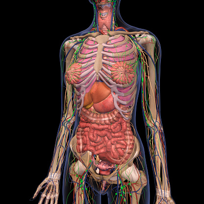
Human Anatomy Of Female Chest And Abdomen 2 Stock Photo ... from media.istockphoto.com The four anatomical regions of the abdomen are known as quadrants. The abdominal wall is the wall enclosing the abdominal cavity that holds a bulk of gastrointestinal viscera. Learn about its function, parts, abdominal conditions, and more. The diaphragm forms the upper surface of. Divided into 9 regions by two vertical and two horizontal imaginary planes divided into 4 quadrants by single vertical and horizontal imaginary plane. These pelvic organs are covered by the same elastic peritoneum membrane that covers most of the the abdominal cavity is the largest in the body. Abdominal wall anatomy that is clinically pertinent to the surgeon, focusing primarily on the structures of the anterior abdominal wall, will be reviewed. Abdominal soft tissue planes and visceral surfaces are visible on abdominal.
Start your review of anatomy of the chest, abdomen, and pelvis.
Abdominal cavity, largest hollow space of the body. These help bind the muscles together. A neonate with an acute abdomen usually presents with vomiting, constipation and distention of the belly. See more of dr mitch's anatomy videos. The abdomen (colloquially called the belly, tummy, midriff or stomach) is the part of the body between the thorax (chest) and pelvis, in humans and in other vertebrates. Divided into 9 regions by two vertical and two horizontal imaginary planes divided into 4 quadrants by single vertical and horizontal imaginary plane. Abdominal soft tissue planes and visceral surfaces are visible on abdominal. The resonant tone produced by percussion over the anterior chest wall will be somewhat less. Gsi asked questions about the visualize the abdomen as a completely empty shell. The abdomen (commonly called the belly) is the body space between the thorax (chest) and pelvis. Learn about chest and abdomen anatomy with free interactive flashcards. The wall of the abdomen is a muscular structure. Of sectional anatomy, computed tomography and magnetic resonance imaging:
Of sectional anatomy, computed tomography and magnetic resonance imaging: anatomy of chest. Use the mouse scroll wheel to move the images up and down alternatively use the tiny arrows (>>) on both side of the image to move the images.